Attention Members: Please avoid clicking on any unsolicited emails with attachments. If you have any concerns, please contact us at info@veteducation.com.au for clarification.
Attention Members: Please avoid clicking on any unsolicited emails with attachments. If you have any concerns, please contact us at info@veteducation.com.au for clarification.
Dr. Philip R Judge BVSc MVS PG Cert Vet Stud MACVSc (Vet. Emergency and Critical Care; Medicine of Dogs)
Laryngeal paralysis is defined as complete or partial failure of the arytenoid cartilages and vocal folds to abduct during inspiration1. Laryngeal paralysis may result from dysfunction of laryngeal muscles, recurrent laryngeal, or vagus nerves, or cricoarytenoid ankylosis. Of these potential causes, neurological causes are the most common1.
Aetiology
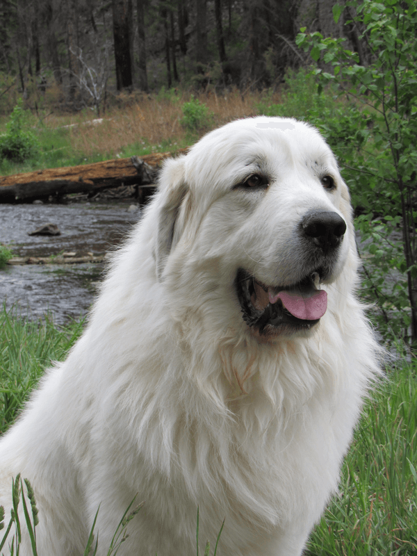
Pyrenean Mountain Dogs have an autosomal recessive inheritance of a polyneuropathy that can cause laryngeal paralysis, among other axonopathies
A congenital form of laryngeal paralysis occurs in certain breeds, such as Siberian huskies, bull terriers, White-coated German Shepherds and Bouvier de Flandres1-3. A hereditary predisposition has also been identified in Alaskan malamutes, and crosses of these breeds2,3, A laryngeal paralysis-polyneuropathy complex has been identified in Dalmatians, Rottweilers, Leonberger and Pyrenean mountain dogs2,3,.
More frequently, laryngeal paralysis is acquired, with proposed causes including trauma, cervical masses and neuromuscular diseases1-3. In most cases, however, the cause remains undetermined and these have traditionally been classified as idiopathic. However, many of these dogs may develop systemic neurological signs within 12 months or so following diagnosis of laryngeal paralysis. Nerve and muscle analysis of these patients suggest the condition is consistent with a progressive, generalised polyneuropathy. The description “Geriatric-Onset Laryngeal Paralysis Polyneuropathy” or GOLPP has been proposed as a more accurate description of laryngeal paralysis in these dogs2,3.
Idiopathic dysfunction or degeneration of the recurrent laryngeal nerve appears to be the most common cause of laryngeal paralysis in dogs1. In this condition, the neurological supply to the dorsal cricoarytenoid muscle by the recurrent laryngeal nerve is abnormal, and results in atrophy of the muscle tissues, and a failure of abduction of the arytenoid cartilage at inspiration. Typically, this results in the presence of a partial upper airway obstruction and symptoms of upper respiratory stridor during inspiration1.
As outlined above, laryngeal paralysis is a disease of diverse aetiology. However, in most dogs, an underlying cause is not identified. These cases are referred to as idiopathic laryngeal paralysis. The following table lists some of the known or suspected causes of laryngeal paralysis, along with commonly associated breed representations in the disease.
Causes of Laryngeal Paralysis in Dogs
Congenital
Bouvier de Flandres
Autosomal dominant inheritance
Dogs usually affected at <1 year of age
Bull Terriers
Wallerian degeneration of recurrent laryngeal nerve
Abnormal nucleus ambiguous
Affected at <1 year
Pyrenean Mountain Dogs
Polyneuropathy – concurrent megaoesophagus and distal axonal degeneration of peripheral nerves
As mentioned above, laryngeal paralysis results in failure of abduction of arytenoid cartilages, and consequently also, the vocal folds, during inhalation, causing upper airway obstruction. Dogs typically present with noisy inspiratory respiration, with varying degrees of exercise intolerance. Advanced or severe cases of laryngeal paralysis display symptoms of near or complete airway obstruction during inhalation. Other symptoms are outlined in the table below:
Clinical Signs of Patients with Laryngeal Paralysis or Upper Airway Disease
General Symptoms
Inspiratory stridor
Exercise intolerance
Decreased activity level
Voice change
Ptyalism
Cough
Dysphagia
Severe Symptoms
Cyanosis
Collapse
Gagging
Retching
Syncope
Vomiting
Death
When accompanied or exacerbated by exercise, some patients may also show symptoms of hyperthermia, ataxia, collapse and even death. These symptoms typically result from an increase in respiratory rate and effort, increasing turbulence of airflow through the larynx, inflammation and swelling of the laryngeal mucosa, resulting in oedema and further airway obstruction, which can be life-threatening1-3.
Laryngeal paralysis occurs in large breed dogs more commonly than small breed dogs, and occurs 2-4 times more frequently in males than females. Inherited causes of laryngeal paralysis usually result in symptoms in affected dogs at less than 1 year of age; while acquired laryngeal paralysis usually occurs in dogs during middle age or older1.
Diagnosis
The diagnosis of laryngeal paralysis is typically made using a combination of supportive clinical signs, followed by direct or laryngoscopic visualization of laryngeal cartilages during inspiration and expiration under a light (stage 2-3) of anaesthesia. However, a complete patient evaluation is necessary to assess the patient for the presence of concurrent or underlying disease2,3. For example, aspiration pneumonia is a common complication of bilateral laryngeal paralysis, making thoracic radiography a necessary evaluation tool. In addition, consideration must be given to other causes of airway obstruction, including brachycephalic airway syndrome, laryngeal collapse, tracheal collapse, trauma, and airway obstruction with foreign material, soft tissue swelling or neoplasia1-4.
The following table lists the most common diagnostic tests appropriate for patients with laryngeal paralysis1-4
Diagnostic Test
Rationale
Physical Examination
Evaluation of patient general health
CBC/Serum Biochemistry
Evaluation of general body function
Urinalysis
Evaluation of general body function
Thyroid Function Tests
Possible association between hypothyroidism and occurrence of laryngeal paralysis (30% of laryngeal paralysis dogs have hypothyroidism)
Thoracic Radiography
Evaluation of pulmonary parenchyma (aspiration pneumonia); evaluation of esophagus (megaoesophagus)
Oesophogram
Evaluation of megaoesophagus in patients with dysphagia or vomiting
Ultrasonography of the Larynx
Can accurately identify laryngeal paralysis by assessing cuneiform process and arytenoid cartilage movement
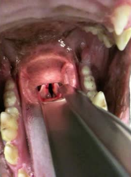
Direct visualization of Larynx
No movement or paradoxical movement of arytenoid cartilages is seen under light plane of general anaesthesia
Laryngeal examination under a light plane of anaesthesia is required to provide a definitive diagnosis or laryngeal paralysis, and to rule out other laryngeal abnormalities1-5. Appropriate laryngeal evaluation requires general anaesthesia, as mentioned in the table above.
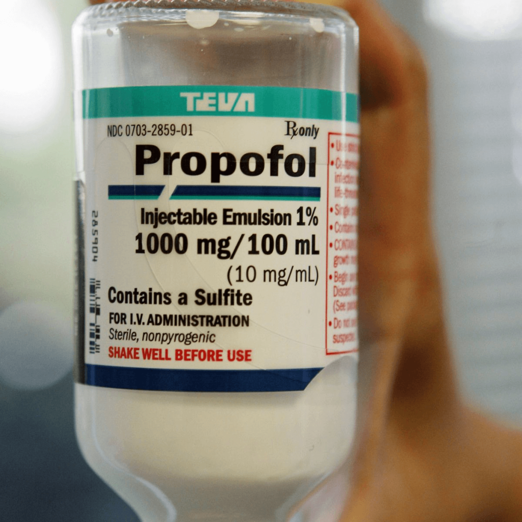
Anaesthetic agents that have been studied for accurate evaluation of the larynx include thiopentone, propofol, Alfaxalone, and ketamine. Additionally, the inclusion of premedication with agents including opioids, acepromazine and alpha-2 agonists has been studied2,3,5,6. A brief overview of the use of anaesthetic agents is outlined below:
Propofol is commonly used as an induction agent to visualise laryngeal movement under light anaesthesia
Premedication
Dexmedetomidine: when used alone, or in combination with either butorphanol or hydromorphone, dexmedetomidine does not appear to inhibit laryngeal movement5,7.
Butorphanol: Butorphanol has been evaluated as a sole pre-anaesthetic medication in 2 studies, at doses ranging from 0.2-0.5 mg/kg. Laryngeal movement was preserved in both studies, when anaesthesia was induced with either propofol or thiopenone5,6.
Acepromazine and butorphanol: Addition of acepromazine to butorphanol pre-anaesthetic protocol resulted in lack of arytenoid movement in 50% of dogs subsequently anaesthetised with either propofol or alfaxalone5.
Acepromazine: Acepromazine, when used as the sole pre-anaesthetic agent, resulted in significant decreases in laryngeal movement, regardless of induction agent used5,6.
Methadone: a single study evaluated the use of methadone in combination with acepromazine, and found a 15% loss of detectable arytenoid motion when anaesthesia was induced with propofol or alfaxalone5.
Induction agents
Propofol: six out of 8 studies evaluating propofol found no significant differences between propofol and other induction agents, including thiopentone, alfaxalone, and diazepam/ketamine. However, two studies found significantly poorer laryngeal function following induction in non-premedicated dogs, resulting in false-positive diagnoses of laryngeal paralysis. Overall, propofol shows high variability in laryngeal movement following induction, despite the widespread use of this induction agent for laryngeal function assessment. Dose, technique (slow administration vs. bolus administration), and presence of post-induction apnoea may be important in interpreting the results of examination of the patient larynx when using propofol5,6.
Alfaxalone: alfaxalone induction, when used in premedicated or un-premedicated dogs, resulted in similar laryngeal function to that observed in comparative studies with propofol and thiopentone. Interestingly, low-dose alfaxalone (1.9 mg/kg induction dose) appeared to result in less laryngeal movement preservation than higher induction doses (2.6 mg/kg)5.
Thiopentone: thiopentone has been extensively studied as an induction agent for evaluation of laryngeal function. Several comparative studies have found no statistical difference in laryngeal movement when thiopentone is compared to alfaxalone, propofol, propofol/diazepam, and ketamine/diazepam5.6.
Diazepam/ketamine: studies evaluating the use of diazepam/ketamine for laryngeal assessment report divergent results, with some reporting similar laryngeal function to thiopentone, and propofol; but with others reporting absent laryngeal movement scores5,6.
Isoflurane mask-induction: mask induction with isoflurane has been evaluated, and revealed retention of laryngeal movement until jaw tone relaxation occurred. High doses of acepromazine were noted to be required in order to facilitate mask induction5.
Administration of doxapram can increase force and depth of respiratory effort under anaesthetic, exacerbating intrinsic laryngeal motion by up to 60%, and should be used where routine laryngeal evaluation results in equivocal diagnosis.
Doxapram has been evaluated as a respiratory stimulant in two-thirds of studies, and successfully increased laryngeal motion in 75% of these studies. It should be noted that in one study, there was no increase in laryngeal movement in non-premedicated dogs following doxapram administration, whereas in another study, doxapram was effective in stimulating laryngeal function. Doxapram was effective in stimulating laryngeal movement in premedicated dogs, including some patients who previously lacked arytenoid motion. Interestingly, in one study, laryngeal function was not observed in 50% of dogs induced with alfaxalone5.The goal of anaesthesia during the evaluation of laryngeal function in dogs is aimed at providing adequate sedation, while maintaining laryngeal motion and reflexes mostly intact (Stage 3, Plane 2 anaesthesia). Based on a review of anaesthetic regimes for laryngeal assessment, there does not appear to be sufficient evidence to clearly recommend one single anaesthetic regime for the evaluation of laryngeal function in dogs. However, current evidence suggests that premedication maintains overall adequate laryngeal motion in dogs and provides better conditions for laryngeal examination than achieved with sole use of induction agents5-7. Any negative impact on arytenoid motion caused by premedication may be overcome with doxapram. Using either propofol or alfaxalone alone is not recommended for the evaluation of arytenoid motion. Doxapram is effective in differentiating normal dogs from dogs with laryngeal paralysis5,8,9.
Treatment
Emergency Treatment
Goals in emergency management of symptoms resulting from suspected or confirmed laryngeal paralysis include1-3
Improvement of ventilation
Reducing laryngeal oedema
Reducing patient stress
These goals are typically achieved by
Providing flow-by oxygen supplementation, or trans-tracheal oxygen catheter1-3
Administration of short-acting intravenous corticosteroids – methyl-prednisolone sodium succinate 5-10 mg/kg IV once1-3
Administration of sedatives – acepromazine 0.01-0.02 mg/kg IV either alone or in combination with butorphanol 0.1-0.2 mg/kg IV1-3
Cooling – many patients suffering from airway obstruction can become hyperthermic either due to poor thermoregulation, environmental conditions, or both. Cooling should take place using a combination of cool water flow over the animal, and by maximizing airflow over the patient using a fan to increase convective heat loss. Cool water enemas may also be employed if required. Active cooling efforts should cease once rectal temperature falls to 39.5 degrees Celsius1-3
Patients who present in severe respiratory distress with airway obstruction, or in those who fail to improve with aforementioned efforts, may require tracheal intubation with endotracheal tube, or emergency tracheotomy – should be performed, in order to by-pass the obstructed larynx1-3.
Trans-laryngeal percutaneous arytenoid lateralization is currently under investigation as an alternative to temporary tracheostomy for dogs in severe respiratory distress from laryngeal paralysis, and involves placement of a mattress suture through one arytenoid cartilage, with the suture exiting the skin ventral to the jugular vein10,11.
Surgical Treatment
Surgical management is recommended for patients with laryngeal paralysis that have moderate-to-severe signs of respiratory distress1-3. The goal of treatment is to widen the glottis, while maintaining some protection against aspiration of saliva and food material into the airways. Many surgical techniques are described for the management of laryngeal paralysis, with unilateral arytenoid lateralization being the most preferred. Outcomes of these techniques are summarized below:
Vocal fold excision12
Involves excision of vocal folds to widen the ventral glottis.
Can be effective in mild cases of airway compromise
May not reduce airway resistance
Glottic stenosis occurs in up to 20% of cases, which can be difficult to manage
Partial Arytenoidectomy12
Involves removal of the corniculate process of the arytenoid cartilage, enlarging the dorsal aspect of the glottis
Associated with serious complications (including death) in 50% of cases in the post-operative period
Re-innervation of the larynx
Muscle-nerve pedicle transposition using phrenic nerve branches; or hypoglossal nerve branches
Improvement in function may take 5-11 months to occur
May be useful in mild to moderately affected dogs
Modified castellated laryngofissure13
Involves castellated laryngofissure, vocal fold resection, and bilateral arytenoid lateralization
Tidal breathing flow volume loop values were normal in 7 of 10 dogs evaluated within 5 to 189 days after surgery
The procedure provided successful long-term relief of upper airway obstruction associated with bilateral laryngeal paralysis
Unilateral arytenoid lateralization3,14
Several techniques described
Suturing cricoid cartilage to the muscular process of the arytenoid cartilage. This technique mimics the directional pull of the cricoarytenoid
Suture placement from the muscular process of the arytenoid cartilage to the caudodorsal aspect of the thyroid cartilage. This technique pulls the arytenoid cartilage laterally rather than rotating it and increases the area of the rima glottis to a lesser degree than the cricoarytenoid suture.
Less invasive techniques have been described, that involve less dissection of the thyro-pharyngeus muscle and preservation of the crico-pharyngeus muscle, cricothyroid articulation, and sesamoid bands, in an effort to preserve laryngeal structure. No differences in complication rates or survival times were noted with this technique in a recent study
Complications and Prognosis
Aspiration pneumonia is the most common complication in patients with laryngeal paralysis, occurring in 10% to 21% of dogs undergoing unilateral arytenoid lateralization. Although aspiration pneumonia is most likely in the first few weeks following surgery, these dogs are at risk of this complication for the rest of their lives3.
Factors increasing risk of aspiration pneumonia include1-3:
Oesophageal dysfunction
Progression of neurological signs (GOLPP)
Temporary tracheostomy placement
Concurrent neoplasia
Post-operative megaoesophagus
Post-operative opioid administration
The use of gastrointestinal motility modifiers has been suggested to reduce the risk of postoperative aspiration pneumonia, given a large number of dogs with laryngeal paralysis have evidence of gastro-oesophageal reflux15.
Metoclopramide increases lower oesophageal sphincter tone and has been shown to reduce gastroesophageal reflux under anesthesia in normal dogs; however, a prospective multicenter clinical trial in dogs with laryngeal paralysis undergoing unilateral arytenoid lateralization found that perioperative administration of metoclopramide did not affect the incidence of aspiration pneumonia in the short-term postoperative period16.
Cisapride has been suggested for use to increase lower oesophageal sphincter tone17. A retrospective evaluation of a cisapride suggested a positive effect on the incidence of postoperative aspiration pneumonia, but definitive conclusions could not be drawn because of low numbers of dogs in the study18.
Progressive development of neurological signs can also affect outcome. In one study, dogs with neurologic comorbidities had a higher risk of complications following surgical correction of laryngeal paralysis19.
In the absence of surgical complications, unilateral arytenoid lateralization results in reduced respiratory distress and stridor and improved exercise tolerance. Survival rates are reported as 93.6% at 1 year, and at 84.4% at 4 years post surgery. Development of post-operative aspiration pneumonia reduces survival rates to 83.1% at 1 year, and 51.5% at 3 years20.
Conclusion
larynx of a dog
Laryngeal paralysis in dogs is a serious condition. Acute emergency presentation is often complicated by marked airway oedema, and may be accompanied by life-threatening heat stress, shock, and airway compromise. Acute medical therapy is necessary in these patients, including emergency airway access in some patients.
Surgical therapy is indicated in most patients and can result in good quality of life. Aspiration pneumonia is the most common complication of therapy. In some patients, progressive neuropathy may occur, and adversely affect outcome.
References:
Monnet E, Tobias KM. Larynx. In: Tobias KM, Johnston SA, editors. Veterinary surgery small animal. St Louis (MO): Elsevier; 2012. p. 1724.
MacPhail C. Laryngeal disease in dogs and cats. Veterinary Clinics: Small Animal Practice. 2014 Jan 1;44(1):19-31.
MacPhail CM. Laryngeal Disease in Dogs and Cats: An Update. Veterinary Clinics: Small Animal Practice. 2020 Mar 1;50(2):295-310.
Radlinsky, W. (2009). Comparison of Three Clinical Techniques for the Diagnosis of Laryngeal Paralysis in Dogs. Veterinary Surgery, 38(4), 434–438.
Ranninger E, Kantyka M, Bektas RN. The Influence of Anaesthetic Drugs on the Laryngeal Motion in Dogs: A Systematic Review. Animals. 2020 Mar;10(3):530.
Jackson AM, Tobias K, Long C, Bartges J, Harvey R. Effects of various anesthetic agents on laryngeal motion during laryngoscopy in normal dogs. Veterinary Surgery. 2004 Mar;33(2):102-6.
DeGroot WD, Tobias KM, Browning DC, Zhu X. Examination of laryngeal function of healthy dogs by using sedation protocols with dexmedetomidine. Veterinary Surgery. 2020 Jan;49(1):124-30.
Radkey DI, Hardie RJ, Smith LJ. Comparison of the effects of alfaxalone and propofol with acepromazine, butorphanol and/or doxapram on laryngeal motion and quality of examination in dogs. Veterinary anaesthesia and analgesia. 2018 May 1;45(3):241-9.
Tobias KM, Jackson AM, Harvey RC. Effects of doxapram HCl on laryngeal function of normal dogs and dogs with naturally occurring laryngeal paralysis. Veterinary anaesthesia and analgesia. 2004 Oct 1;31(4):258-63.
Hardie RJ. Translaryngeal percutaneous arytenoid lateralization technique in a canine cadaveric study. Journal of Veterinary Emergency and Critical Care. 2016 Sep;26(5):659-63.
Sample SJ, Hardie RJ, Stein J, Webb J. Evaluation of Translaryngeal Percutaneous Arytenoid Lateralization (TPAL) in dogs with experimentally created laryngeal paralysis. Research in veterinary science. 2018 Apr 1;117:239-45.
Ross JT, Matthieson DT, Nonoe KE, Scavelli TA. Complications and long‐term results after partial laryngectomy for the treatment of idiopathic laryngeal paralysis in 45 dogs. Veterinary Surgery. 1991 May;20(3):169-73.
Smith MM, Gourley IM, Kurpershoek CJ, Amis TC. Evaluation of a modified castellated laryngofissure for alleviation of upper airway obstruction in dogs with laryngeal paralysis. Journal of the American Veterinary Medical Association. 1986 Jun 1;188(11):1279-83.
MacPhail CM, Monnet E. Outcome of and postoperative complications in dogs undergoing surgical treatment of laryngeal paralysis: 140 cases (1985–1998). Journal of the American Veterinary Medical Association. 2001 Jun 1;218(12):1949-56.
Tarvin KM, Twedt DC, Monnet E. Prospective controlled study of gastroesophageal reflux in dogs with naturally occurring laryngeal paralysis. Veterinary Surgery. 2016 Oct;45(7):916-21.
Milovancev M, Townsend K, Spina J, et al. Effect of metoclopramide on the incidence of early postoperative aspiration pneumonia in dogs with acquired idiopathic laryngeal paralysis. Vet Surg 2016;45:577–81.
Kempf J, Lewis F, Reusch CE, et al. High-resolution manometric evaluation of the effects of cisapride and metoclopramide hydrochloride administered orally on lower esophageal sphincter pressure in awake dogs. Am J Vet Res 2014;75: 361–6.
Ogden J, Ovbey D, Saile K. Effects of preoperative cisapride on postoperative aspiration pneumonia in dogs with laryngeal paralysis. J Small Anim Pract 2019;60:183–90.
Bookbinder LC, Flanders J, Bookbinder PF, et al. Idiopathic canine laryngeal paralysis as one sign of a diffuse polyneuropathy: an observational study of 90 cases (2007-2013). Vet Surg 2016;45:254–60.
Bookbinder LC, Flanders J, Bookbinder PF, et al. Idiopathic canine laryngeal paralysis as one sign of a diffuse polyneuropathy: an observational study of 90 cases (2007-2013). Vet Surg 2016;45:254–60.
Learn all about the the basic principles of genetic testing, sample collection, reliable database information, test result interpretation, and recommendations for clients.
This article will explore the presentation, pertinent pathophysiology, patient selection, treatment considerations and outcomes of medical management of canine pyometra.
This short review will focus on the concept of oxygen toxicity, and its relevance in the treatment of the trauma patient - many of whom require and are given oxygen therapy as part of their treatment regime.