Attention Members: Please avoid clicking on any unsolicited emails with attachments. If you have any concerns, please contact us at info@veteducation.com.au for clarification.
Attention Members: Please avoid clicking on any unsolicited emails with attachments. If you have any concerns, please contact us at info@veteducation.com.au for clarification.
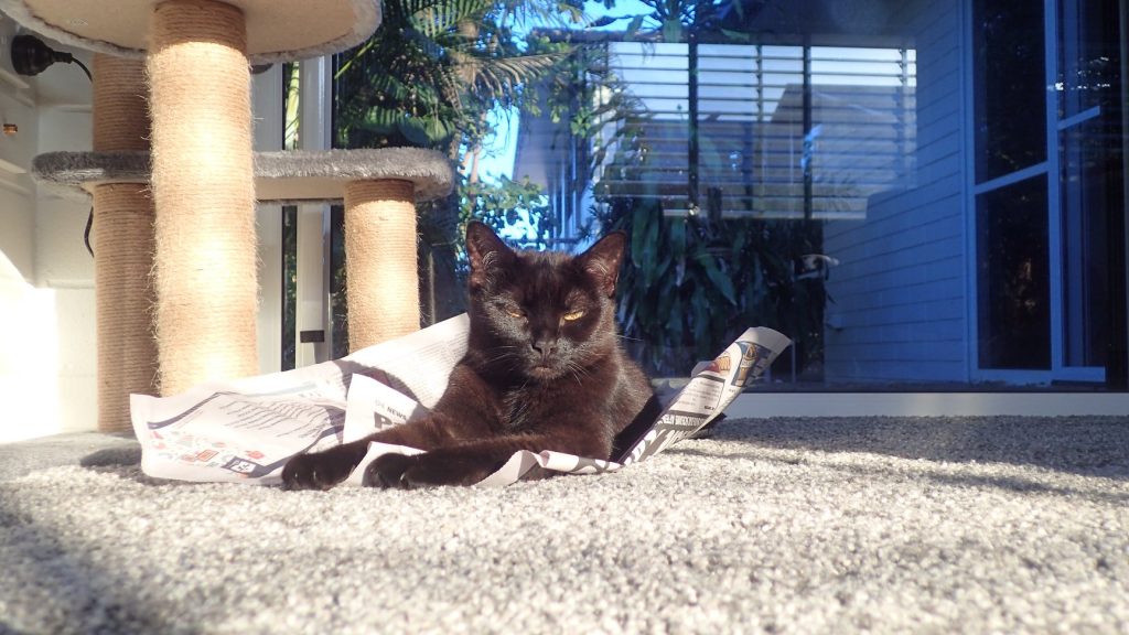
Listen to Dr. Philip Judge talking about Feline Circulatory Shock
Why Talk About Shock in Cats?
Despite the large numbers of studies investigating both physiology and treatment of shock syndromes in both people and dogs, there is a relative paucity of studies on this topic in cats1. Let’s begin with describing shock in cats. The clinical syndrome of shock in cats2 has been characterised by 3 things…
Hypotension
Bradycardia
Hypothermia
This contrasts with the situation in dogs, where we tend to see – at least in hypovolaemic shock – a transition from stage 1 shock – in which hypertension, tachycardia and bounding pulses are seen – to stage 2 shock – in which tachycardia, falling blood pressure, and weaker pulses are seen – to end-stage shock, characterised by tachycardia or bradycardia, hypothermia and low blood pressure1.
Image Courtesy: Batman
Why the difference? What is going on in cats, that is so different to dogs?
The answer is multifactorial – but is thought to principally lie in the acute physiological responses to hypovolaemia or haemorrhage in the cat, that differ from those in the dog.
To help explain this, let’s briefly review the response to hypovolaemia in the dog1-3—
Stage 1 shock (compensatory shock)
Acute hypovolaemia results in activation of the sympathetic nervous system, leading to
Tachycardia
Positive inotropy
Vasoconstriction in gastrointestinal, liver, renal and skin vasculature
Vasodilatation in heart, lungs, brain and muscle
This physiological response is manifested clinically with tachycardia, bounding pulses, heightened mentation and pink mucous membranes with rapid capillary refill time.
Stage 2 shock
Compensatory shock is very short-lived in most patients, and leads to ATP-depletion in vasoconstricted tissues, with progressive vasodilatation in the gastrointestinal tract, skin, liver and kidneys – massively increasing vascular volume. Clinically, this is manifested as a patient with tachycardia, weak pulses, pale mucous membranes, and prolonged capillary refill time.
Stage 3 shock
As stage 2 shock progresses, vascular endothelial damage results from tissue hypoxia caused by poor tissue perfusion. Coagulation activation ensues, followed by widespread thrombosis of micro-vasculature. Organ failure results. Clinically, this is manifested as bradycardia, hypothermia, organ dysfunction (vomiting, diarrhoea, acute kidney failure, arrhythmias, etc.) and death.
Whilst stage 1 shock may occur in cats, it is rarely seen clinically2. Likewise, the “classic” decompensatory characteristics of stage 2 shock are rarely noted in the cat – with bradycardia and hypotension being the predominant clinical signs2.
Why Are Symptoms of Shock in Cats Different?
The Feline Ventricle
Within the wall of the ventricle, the cat has mechanical and chemical volume receptors that respond to distension
Activation of these mechanoreceptors in hypovolaemia reduces heart rate via a cardiac inhibitory reflex, allowing ventricular contraction only when normal ventricular filling pressures are reached in diastole4.
The Feline Atria
The feline atria have feline-specific type-B atrial volume receptors that respond to atrial distension4
Feline-specific type-B receptors discharge during atrial filling and higher pressures, and lead to increases in heart rate; hypovolaemia reduces these receptor stimulation, with bradycardia being the predominant effect.
In both cats and dogs with hypovolaemia, sympathetic nervous system activity increases, which leads to shunting of blood into cardiac and respiratory circulation, increasing atrial distension. In cats, however, there is an inhibition of the cardiac sympathetic efferent nerve activity following stimulation of type B atrial receptors, as atrial pressures rise.
In hypovolaemia, decreased atrial stretch activity related to action of the atrial volume receptors occurs before carotid sinus and aortic baroreceptors and leads to hypothalamic-mediated increases in vasopressin concentrations, increasing blood volume.
3. Noradrenaline and Hypothermia
Noradrenaline release from sympathetic nerves leads to a centrally-mediated hypothermia5,6
Hypothermia reduces noradrenaline release in the CNS and in the heart, blunting sympathetic nervous system responses.
Parasympathetic Nervous Outflow
Increased parasympathetic outflow and inhibition of sympathetic outflow, occurs by the Benzold-Jarisch reflex (BJ reflex) in hypothermic cats, producing bradycardia and vasodilatation7.
As shock progresses, prolonged tissue hypoxia leads to extensive cellular death, organ failure and eventually, patient death (stage 3 – or end-stage shock)
The Causes of Shock in the Cat
The causes of shock in the cat are broadly2,3—
Hypovolaemic shock
Caused by fluid loss from conditions such as
gastrointestinal losses
Third-space losses
Inadequate fluid intake
Haemorrhage
Trauma
Systemic inflammation
Cardiogenic shock
Caused by cardiac conditions, including
Cardiomyopathy
Myocardial ischaemia
Mitral valve insufficiency
Cardiac arrhythmias
Sepsis
Distributive shock
Caused typically by severe illness or infection, among other causes, including
SIRS
Sepsis
Severe liver disease
Anaphylaxis
Spinal cord injury
Obstructive shock
Results from obstruction to venous return to the heart, or increased afterload. Causes include
Pneumothorax
Venous thrombosis
Cardiac tamponade
Caval syndrome (neoplastic occlusion, heartworm)
Positive-pressure ventilation
Treatment of Feline Circulatory Shock
Aside from basic supportive care, including provision of oxygen therapy, analgesia etc., the treatment of shock in the cat may be divided into four (4) phases1-3, 8—
Phase 1: Determine the type of shock present
Assess the following to determine the type of shock present, as this will assist in directing treatment
History – the presence of trauma, bite wounds, gastrointestinal symptoms, potential toxin exposure, medical history may provide vital information that can inform the veterinary team on possible causes of shock.
Clinical examination – physical examination of the patient may reveal the characteristic signs of shock (bradycardia, hypotension and hypothermia), but may also reveal other signs that may help determine aetiology, including
Hydration status
Abdominal pain
Jaundice
Wounds
Haemorrhage
Cavity fluid accumulation
Pneumothorax or other pleural space disease
Cardiac murmurs, gallop rhythms, jugular pulse etc.
Diagnostic evaluation – diagnostic test selection will vary, depending on the patient signalment and clinical examination, but may include the following
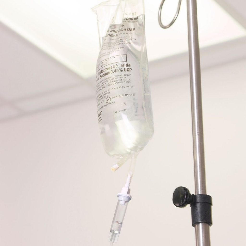
Fluid selection in the treatment of shock in the cat is made based on clinical status, combined with laboratory assessment of hematocrit and plasma protein, along with electrolyte and acid/base analysis.
Crystalloid fluids will distribute within the entire extracellular space following administration – meaning that up to 75% of the administered crystalloid fluid will distribute to the interstitial spaces, leaving as little as 25% within the intravascular space. These fluids are useful in managing acute hypovolaemia – but it is important to note in some diseases, that redistribution of administered fluid into interstitial spaces may lead to patient compromise, e.g., pulmonary contusions – meaning judicious use is necessary.
Colloid fluids are suspensions of large molecular weight compounds (glucose polymer, starches proteins or gels). These large molecules do not readily pass through intact capillary membranes – meaning more of the administered solution will remain within the intravascular space. These fluids can be useful in managing acute hypovolaemia in patients with intact capillary membranes, e.g., acute hypovolaemia. However, starch-based colloids should be avoided in patients with sepsis, systemic inflammation and potentially, patients with significant increases in capillary permeability, as, at least in humans with sepsis, they are associated with increased risk of kidney damage and mortality.
Fluid therapy in most cats initially presenting with shock will involve judicious use of crystalloids, with occasional use of small bolus colloids in non-septic patients to support crystalloid use if required.
Phase 3: Determine resuscitation end-points
Resuscitation end-points provide a set of values (heart rate, blood pressure, temperature etc.) that are targets for resuscitation (so-called “goal-directed resuscitation”), following which, the rate of fluid therapy is reduced to maintenance rates, plus amounts sufficient to correct hydration deficits and ongoing patient fluid loss.
Whilst traditional goal-directed therapy has reduced mortality rates in humans with sepsis and trauma, it is important to individualise patient treatment, so that optimum outcome is achieved9.
Bradycardia, hypothermia and hypotension are associated with worse outcome in cats10.
Appropriate resuscitation end-points in the cat with shock include
Systolic arterial pressure = 100-120 mm Hg
Heart rate: 160-180/min
Normalisation of body temperature. Note that this parameter should be corrected following commencement of fluid resuscitation and should be raised to an initial level of 36.7 degrees over the first 30-40 minutes of resuscitation. Hypotension will begin to resolve as body temperature rises.
In addition, patients with blunt force abdominal trauma, abdominal haemorrhage, pulmonary contusions or traumatic brain injury may benefit from administration of the anti-thrombolytic medication, tranexamic acid.
Phase 4: Administer fluid therapy
Small volume resuscitation is the technique of choice and involves administering 7 ml/kg of isotonic crystalloid fluid over 10 minutes, followed by patient re-evaluation, to determine if subsequent fluid boluses are required. This process is continued for 20-30 minutes initially. A single bolus of isotonic crystalloid may be substituted for a 3 ml/kg bolus of hydroxy-ethyl starch, if desired, in non-septic patients, and in patients without kidney compromise, to prolong the intravascular effectiveness of the crystalloid fluid11.
The Patient that Does Not Respond to Therapy
Patients with circulatory compromise may sometimes fail to respond to the aforementioned treatment2. Failure to respond to therapy to treat shock should prompt investigation to determine the cause. Common causes of persistent circulatory failure in the cat are outlined below:
Inadequate intravascular volume
Blood loss
Third space losses (gastrointestinal tract, body cavity loss, etc.)
Pain
Heart disease
Cardiac tamponade
Heart failure
Arrhythmias
Blood gas or electrolyte abnormalities
Hypoglycaemia
Hypocortisolaemia
Anaemia
Sepsis
Pleural space disease
CNS disease
Knowledge of the causes of poor response to treatment for shock can assist in selection of diagnostic tests to determine the aetiology in a given patient, so that correction of the underlying cause can commence.
Conclusion
Shock in the feline patient presents with a distinct, and separate clinical syndrome to the dog. Understanding the reasons for this, the potential causes, and having a clear outline of how to treat shock in the cat allows timely and appropriate intervention – both of which will reduce mortality and morbidity.
Vet Education (www.veteducation.com.au) provides an extensive array of courses related to veterinary emergency and critical care. If you have any questions, or to stay up-to-date with our current course offerings, visit the website, join our mailing list today, or email info@veteducation.com.
References:
Davis H, Jensen T, Johnson A, Knowles P, Meyer R, Rucinsky R, Shafford H. 2013 AAHA/AAFP fluid therapy guidelines for dogs and cats. Journal of the American Animal Hospital Association. 2013 May;49(3):149-59.
Rudloff E, Kirby R. Feline Circulatory Shock. In August’s Consultations in Feline Internal Medicine, Volume 7 2016 Jan 1 (pp. 843-858). WB Saunders.
Fahim M. Cardiovascular sensory receptors and their regulatory mechanisms. Indian J Physiol Pharmacol 203 47 (2): 124-146
Myers RD, Beleslin DB, Rezvani AH. Hypothermia: role of α1-and α2-noradrenergic receptors in the hypothalamus of the cat. Pharmacology Biochemistry and Behavior. 1987 Feb 1;26(2):373-9.
Kawada T, Kitagawa H, Yamazaki T, Akiyama T, Kamiya A, Uemura K, Mori H, Sugimachi M. Hypothermia reduces ischemia-and stimulation-induced myocardial interstitial norepinephrine and acetylcholine releases. Journal of Applied Physiology. 2007 Feb;102(2):622-7.
Mark AL. The Bezold-Jarisch reflex revisited: clinical implications of inhibitory reflexes originating in the heart. Journal of the American College of Cardiology. 1983 Jan 1;1(1):90-102.
Perner A, Haase N, Guttormsen AB, Tenhunen J, Klemenzson G, Åneman A, Madsen KR, Møller MH, Elkjær JM, Poulsen LM, Bendtsen A. Hydroxyethyl starch 130/0.42 versus Ringer’s acetate in severe sepsis. New England Journal of Medicine. 2012 Jul 12;367(2):124-34.
Roberts JK, Disselkamp M, Yataco AC. Oxygen delivery in septic shock. Annals of the American Thoracic Society. 2015 Jun;12(6):952-5.
Silverstein DC, Wininger FA, Shofer FS, King LG. Relationship between Doppler blood pressure and survival or response to treatment in critically ill cats: 83 cases (2003–2004). Journal of the American Veterinary Medical Association. 2008 Mar 15;232(6):893-7.
Muir WW. A new way to monitor and individualize your fluid therapy plan. Vet Med (2):76-82. 2013.